FIG. 2.
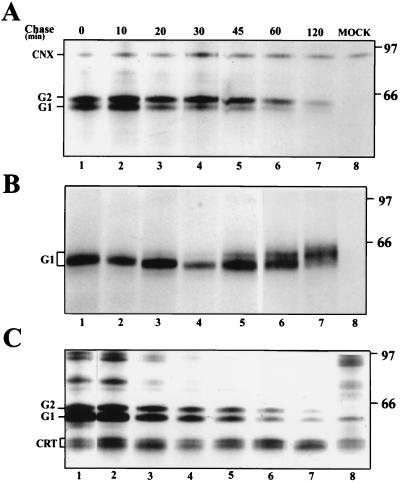
Kinetics of the association of G1 and G2 with calnexin and calreticulin. At 14 h postinfection, BHK21 cells were labeled for 10 min with [35S]methionine and chased for the indicated times. The cells were lysed in 2% CHAPS (A and B) or 1% digitonin (C) and processed for immunoprecipitation with polyclonal anticalnexin (A), anti-G1 (B), or anticalreticulin (C) antisera. The samples in panels A and B are from the same lysate. Immunoprecipitates were analyzed on an SDS–10% polyacrylamide gel under reducing conditions, followed by fluorography. The positions of calnexin (CNX), calreticulin (CRT), G1, and G2, as well as molecular weight markers (in thousands), are indicated. MOCK, immunoprecipitated proteins from uninfected cells.

