. 2024 Jul 22;12(7):e008837. doi: 10.1136/jitc-2024-008837

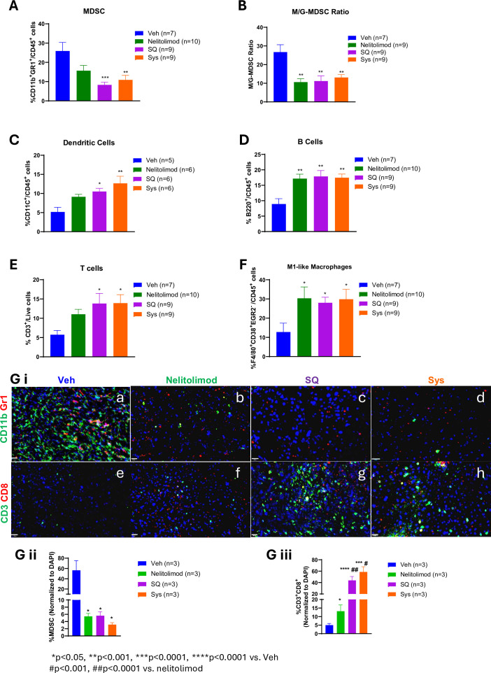
Figure 2. Modulation of liver myeloid and lymphoid compartments by nelitolimod via pressure-enabled drug delivery was preserved in combination with Sys or SQ checkpoint inhibitor. Liver of tumor-bearing mice were harvested 10 days post-treatment. CD45+ cells were isolated from non-parenchymal cells. (A) MDSC cell population (CD11b+Gr1+), (B) monocytic MDSCs (M-MDSC; CD11b+Ly6C+/hiLy6G−/lo), (C) dendritic (CD11c+) cells, (D) B cells (B220+), (E) T cells (CD3+) and (F) M1-like macrophage (F/4/80+CD38+EGR2) were quantified by flow cytometry. (G) i (a–h) Tumors were isolated from each group, OCT-mounted tissues were sectioned, fixed, and stained for CD3 (green), CD8 (red), CD11b (green), and Gr1 (red). (G) ii–iii Quantification of CD11b+Gr1+ MDSCs and CD3+CD8+T cells and from tumors of mice were performed across five fields/mouse and n=3 mice were used per group. Scale (20 µm). Animal data were presented as mean±SEM from and n was mentioned in the individual graph. One-way analysis of variance was performed to determine statistical differences among multiple groups. MDSC, myeloid-derived suppressor cells; M-MDSC, monocytic MDSC; G-MDSC, granulocytic MDSC; OCT, Optimal temperature cutting compound; DAPI, 4′,6-Diamidino-2-phenylindole; SQ, subcutaneous; Sys, systemic; Veh, vehicle.

Figure 2