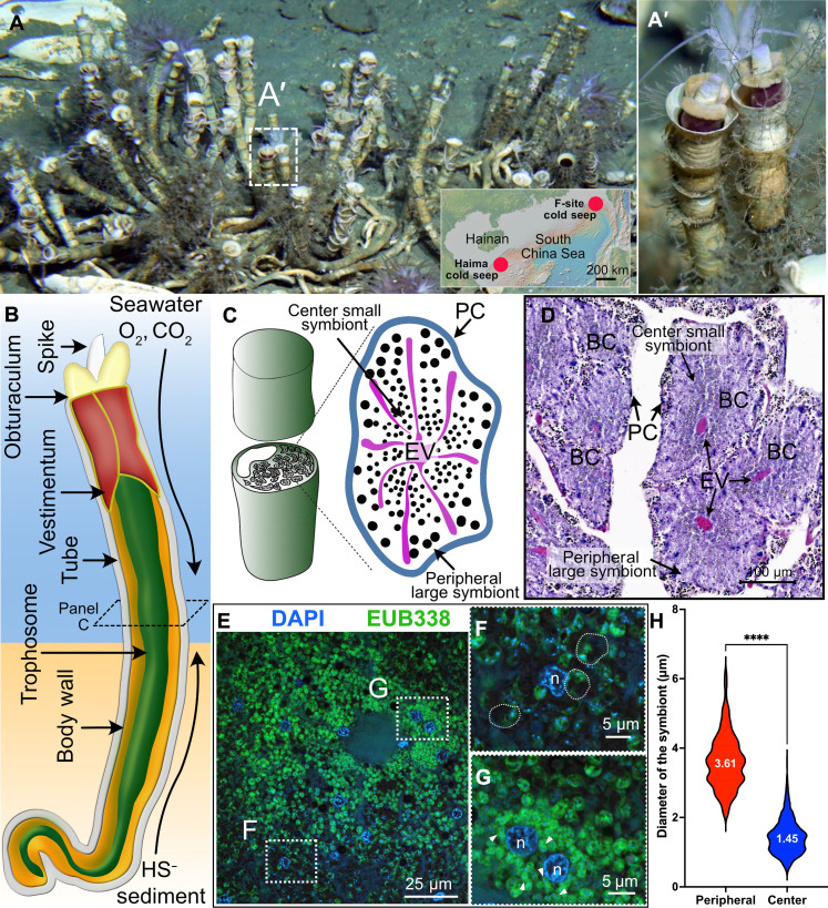
Fig. 1. Natural habitat, gas exchange system, trophosome, and endosymbionts of the deep-sea siboglinid tubeworm P. echinospica.
(A) Photograph of a colony of P. echinospica individuals in the field. Each tube is roughly 30 cm in length. The photo was taken at the Haima cold seep field by the Haima ROV crew. The inserted map shows the regional location map of the two research sites. (B) Diagram of the gas exchange system of P. echinospica. The tubeworm absorbs oxygen and carbon dioxide from the ambient seawater through its plume. It also sucks in sulfide through its posterior region from the sediment. (C) Diagram showing the cross section of each trophosome lobule, with large symbionts located at the lobule periphery and small symbionts at the lobule center. (D) H&E staining of a cross-section of the trophosome of an adult P. echinospia tubeworm. (E) FISH analysis of the symbionts. (F and G) Amplified regions of the periphery and center of a trophosome lobule, demonstrating the morphological variation of the symbionts. The semitransparent dashed lines in (F) outline three typical periphery large symbionts. (H) Statistical analysis of the size variation of bacteriocytes located in the periphery bacteriocytes and center of a lobule (P < 0.001, t test, n-periphery = 449, n-center = 1304).

