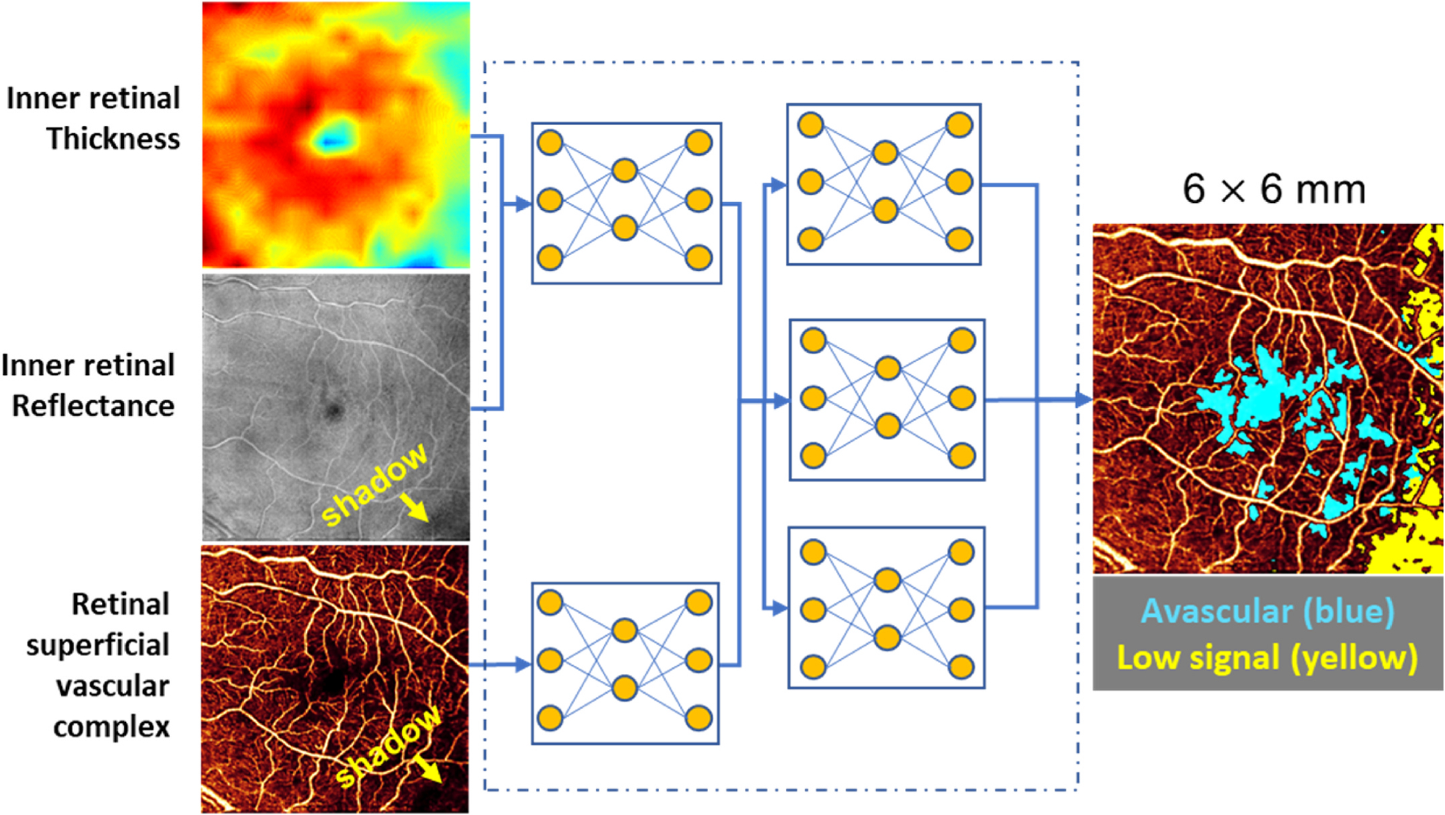
Fig. 10.

Algorithm for distinguishing non-perfusion areas from signal-reduction artifacts on OCTA. With an intelligent combination of structural OCT and OCTA data as the input, the convolutional neural network developed by Guo et al. can accurately distinguish between the real avascular area (blue) and shadow artifacts (yellow). OCT, optical coherence tomography; OCTA, optical coherence tomography angiography. Adapted from Guo, Y. et al., 2019. Biomed Opt Express 10 (7), 3257–3268.
