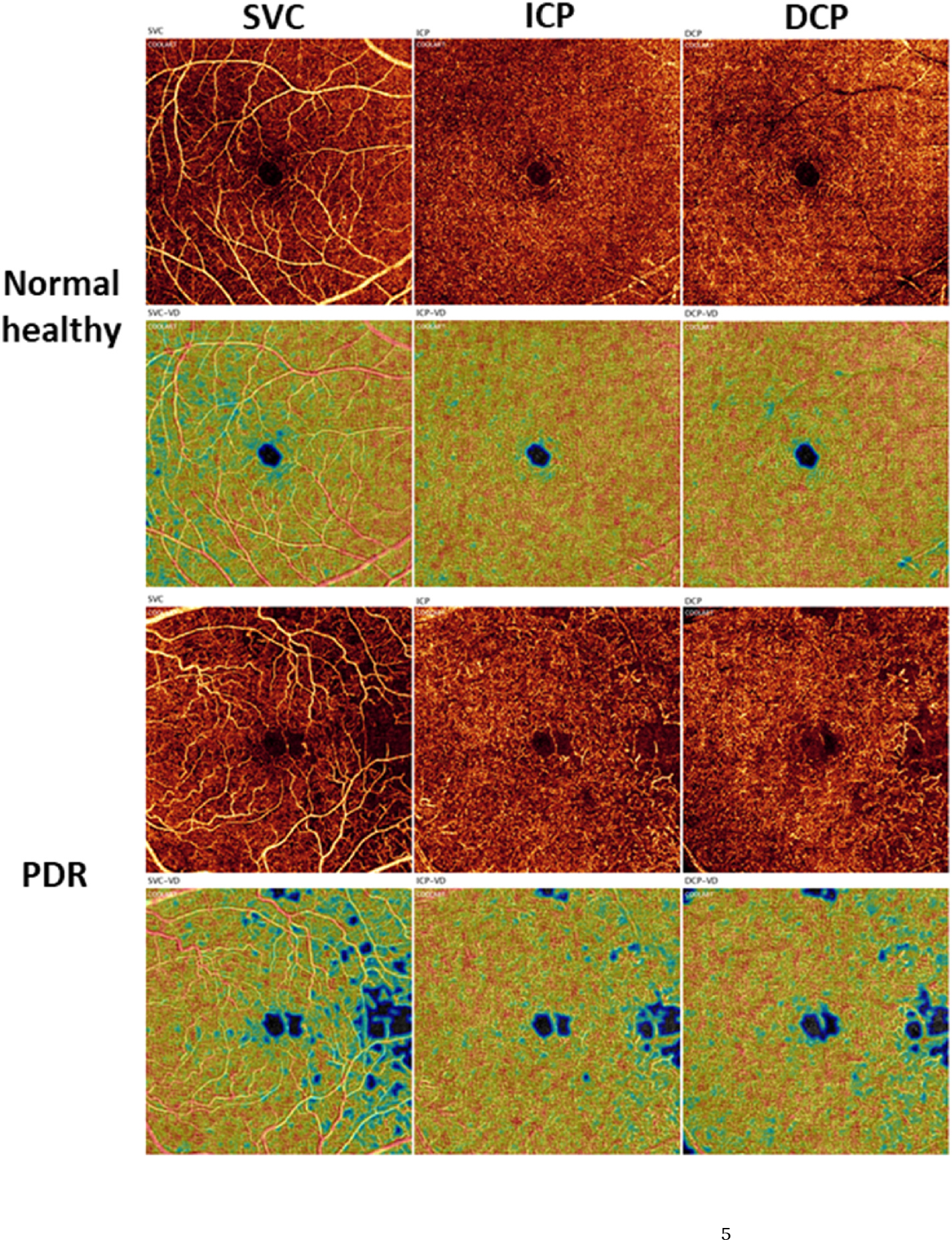
Fig. 3.

Projection-resolved 6 × 6-mm optical coherence tomography angiograms (1st and 3rd rows) and corresponding vessel density heat maps (2nd and 4th rows) of a healthy eye and an eye with PDR in the SVC (1st column), ICP (2nd column), and DCP (3rd column) (previously unpublished data; the published method can be found in (Hagag et al., 2019). Am J Ophthalmol 204, 70–79). DCP, deep capillary plexus; ICP, intermediate capillary plexus; PDR, proliferative diabetic retinopathy; SVC, superficial vascular complex.
