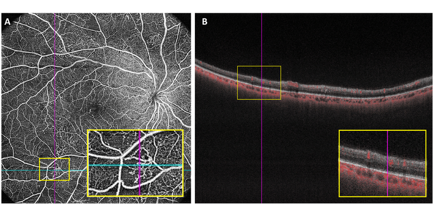
Fig. 6.

Example of IRMAs in one patient with proliferative diabetic retinopathy.(A) 15 × 15 mm Optical coherence tomography angiography image showing IRMA in a full-retinal slab, highlighted in an inset yellow box; (B) B-scan with segmentation lines in the same patient, taken as a cross-section from the blue line in (A), with intraretinal flow highlighted in the inset yellow box. The ILM and hyaloid face are not breached, thus differentiating the IRMA from neovascularization. ILM, inner limiting membrane; IRMA, intraretinal microvascular abnormality.
