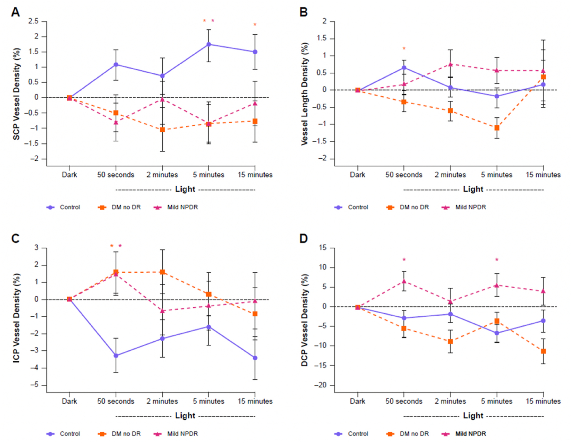
Fig. 7.

Parafoveal vessel density measured using OCTA at different retinal plexuses during ambient light transition for controls, patients with diabetes but no DR, and patients with mild NPDR. (A) Vessel density in the SCP; (B) vessel length density; (C) vessel density in the middle capillary plexus; (D) vessel density in the deep capillary plexus. Statistically significant differences between diabetic and control conditions (p < 0.05) are indicated with an asterisk (*). DCP, deep capillary plexus; DM, diabetes mellitus; DR, diabetic retinopathy; ICP, intermediate capillary plexus; NPDR, non-proliferative diabetic retinopathy; OCTA, optical coherence tomography angiography; SCP, superficial capillary plexus. Adapted from Zhang, Y.S. et al., 2020. J Clin Med 9, 3523.
