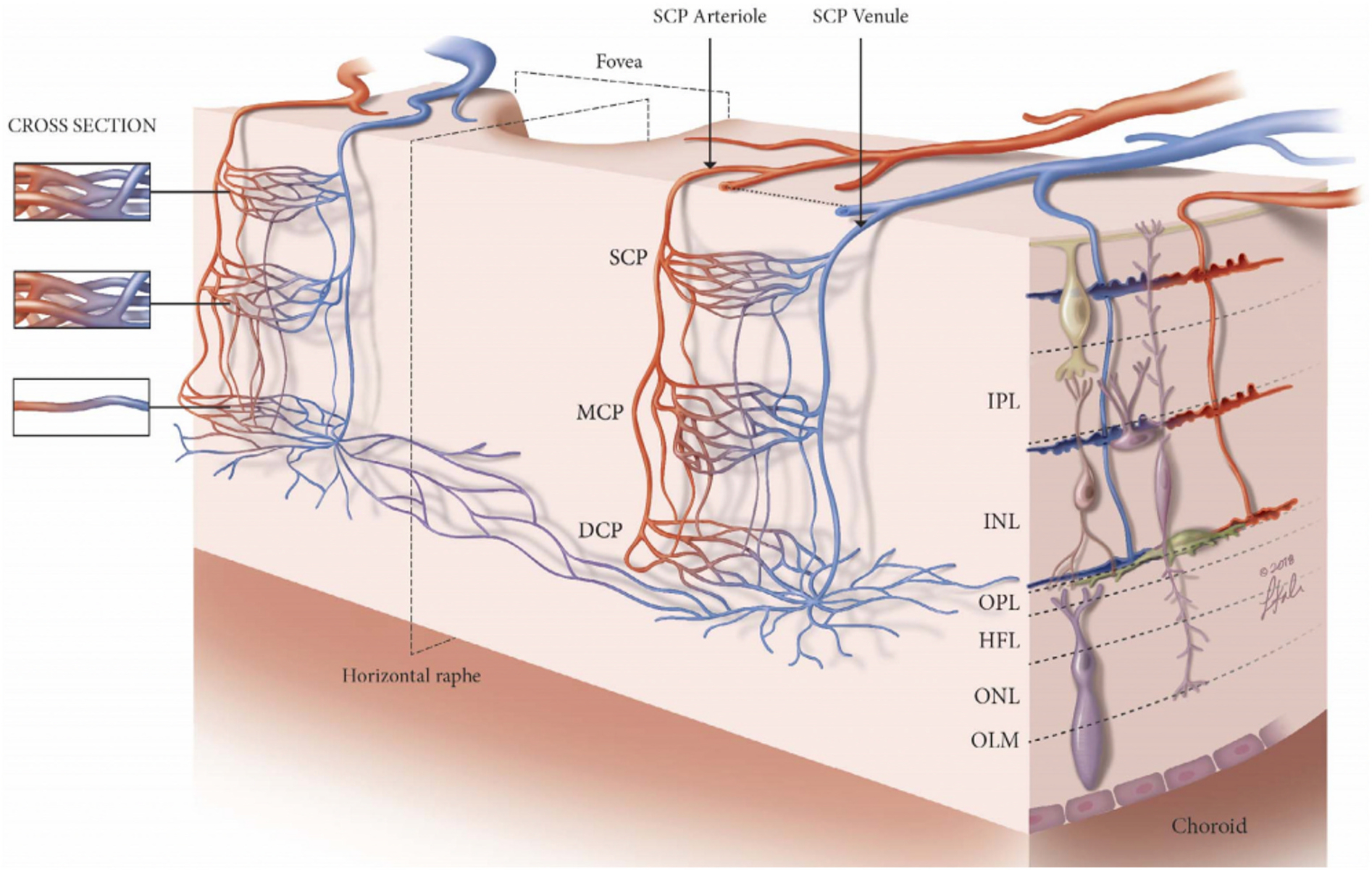
Fig. 1.

Schematic to represent the arrangement of the three retinal plexuses. The superficial capillary plexus (SCP) is located between the retinal ganglion cell layer (RGCL) and the superficial portion of the inner plexiform layer (IPL). The intermediate capillary plexus (ICP), also known as the middle capillary plexus (MCP), starts from the inner border of the IPL to the superficial portion of the inner nuclear layer (INL). The deep capillary plexus (DCP) is distributed across the outer border of the INL. DCP = deep capillary plexus; HFL = Henle’s fibre layer; ICP = intermediate capillary plexus; INL = inner nuclear layer; IPL = inner plexiform layer; MCP = middle capillary plexus; OLM = outer limiting membrane; ONL = outer nuclear layer; OPL = outer plexiform layer; RGCL = retinal ganglion cell layer; SCP = superficial capillary plexuses. Figure courtesy of Nesper PL and Fawzi AA from Human Parafoveal Capillary Vascular Anatomy and Connectivity Revealed by Optical Coherence Tomography Angiography. Invest Ophthalmol Vis Sci. 2018 Aug 1; 59(10):3858–3867. https://doi.org/10.1167/iovs.18-24710.
