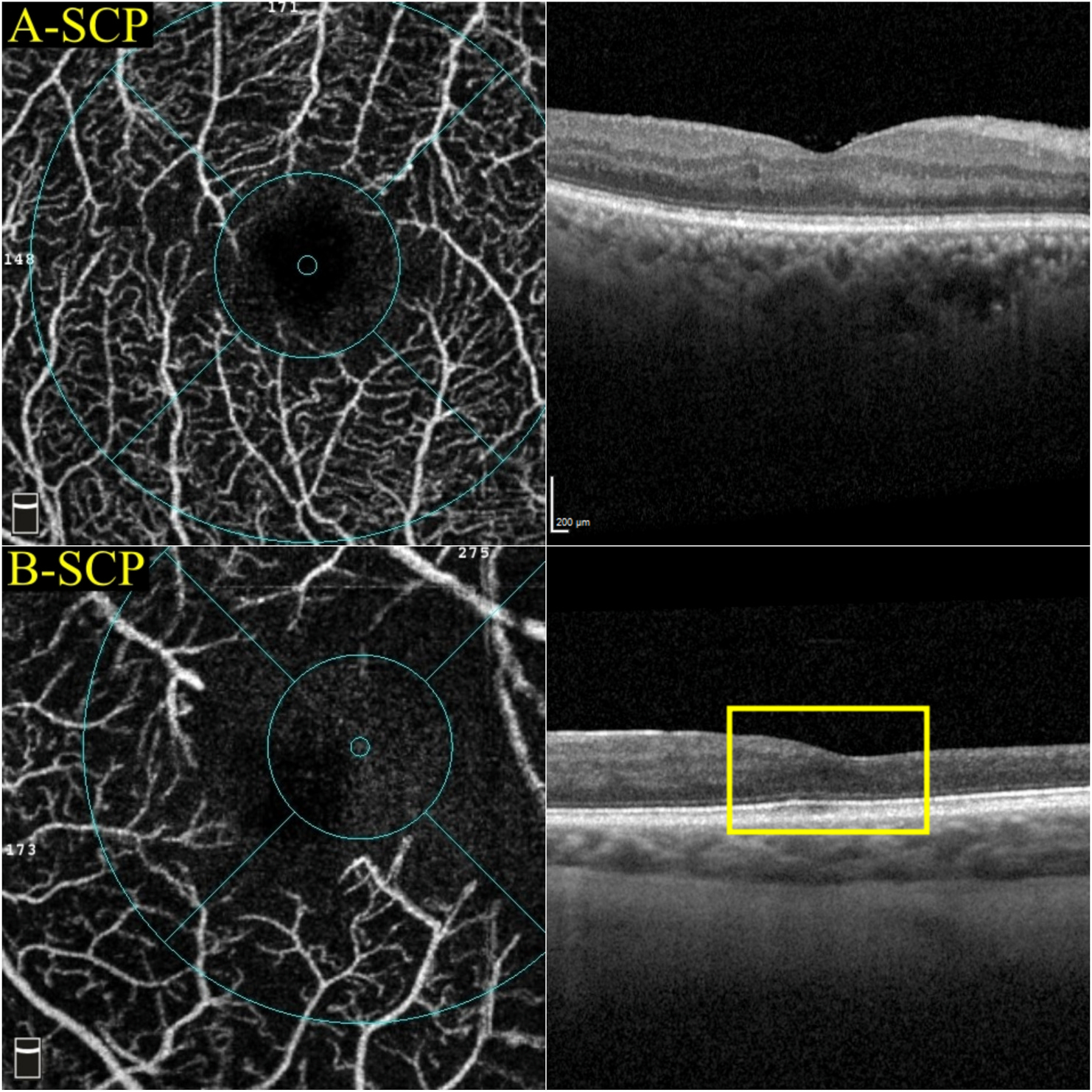
Fig. 12.

A reduced SCP VD on OCTA does not always correspond to the DRIL on OCT. For example, both eye A and eye B had a reduced superficial VD (39% and 25.4%, respectively). However, only eye B presented with DRIL (yellow rectangle). DRIL = disorganisation of retinal inner layers; OCT = optical coherence tomography; OCTA = optical coherence tomography angiography; SCP = superficial capillary plexus; VD = vessel density.
