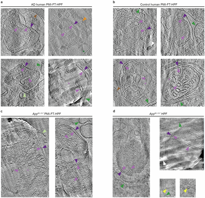
Extended Data Fig. 6. Tomographic slices showing damaged mitochondria in brain tissues that have undergone post-mortem interval and freeze-thaw step.
a, Post-mortem AD brain with PMI and freeze-thaw step that preceded high pressure freezing (PMI-FT-HPF). b, Post-mortem non-demented control brain (Control human PMI-FT-HPF). c, Mouse model of β-amyloidosis brain (AppNL-G-F) prepared with PMI-FT-HPF. d, Mouse model of β-amyloidosis (AppNL-G-F). Sample prepared without post-mortem interval and freeze-thaw step (AppNL-G-F-HPF) (see Leistner et al. for details)30. Dark purple arrowhead, outer mitochondrial membrane. Light purple arrowhead, inner mitochondrial membrane. Light purple asterisk, diluted mitochondrial matrix. Dark green arrowhead, sub-cellular membrane compartment. Light green arrowhead, burst membrane. Orange arrowhead, actin filament. Yellow arrowhead, microtubule. Brown arrowhead, myelin sheath. White arrowhead, knife damage or surface ice contamination. Scale bar, 10 nm.

