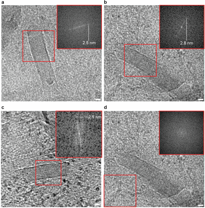
Extended Data Fig. 7. Extracellular cuboidal particles have ordered striations.
a and b, cryoEM images (single tomographic tilt, 2.4 Å pixel size) of an extracellular cuboidal particle in an Aβ plaque (see Extended Data Fig. 3e). Red square, subregion analysed by fast Fourier transform shown in insets with 2.5 nm or 2.8 nm peak, respectively. c, Tomographic slice (9.6 Å voxel size) showing extracellular cuboidal particle. d, Same as b but with no peak in control subregion. Scale bar, 10 nm.

