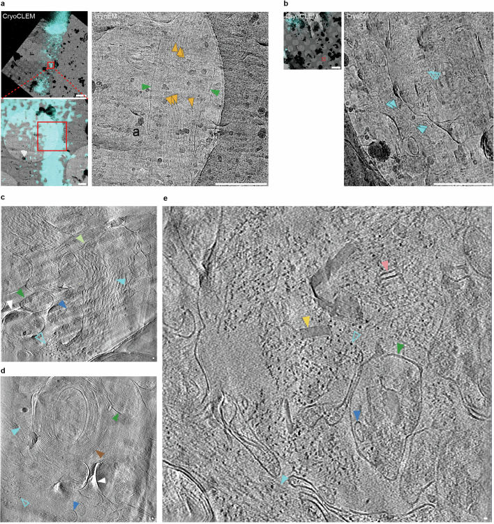
Extended Data Fig. 3. Cryo-CLEM and cryoET of MX04-labelled post-mortem AD brain.
a, CryoCLEM of MX04-labelled tau inclusion within AD post-mortem brain cryo-section. Top left, aligned cryoFM image (cyan, MX04) with cryoEM image. Red rectangle, area shown in close-up. Scale bar, 5 μm. Bottom left, close-up. Red rectangle, region shown, area shown in close-up. Right, close-up. Orange arrowheads, putative tau filaments. Green arrowhead, putative plasma membrane of neurite. Scale bar, 500 nm. See also cryoET data in Extended Data Fig. 8 and Supplementary Video 6). b, CryoCLEM of AD post-mortem brain cryo-section showing unlabelled amyloid deep in the tissue below the depth of MX04 penetration. Left, MX04 (cyan) cryoFM image aligned with cryoEM image showing MX04 only labels top ~15 μm of 100 μm thick tissue biopsy. Red rectangle, region in cryo-section corresponding to 27 μm deep within the tissue biopsy. Scale bar, 5 μm. Right, close-up of left medium magnification cryoEM image showing region from which cryoET data were collected (see e). Cyan arrowhead, putative Aβ fibrils. Scale bar, 500 nm. c-e, Tomographic slices of β-amyloid plaque pathology in post-mortem AD brain. Filled and open cyan arrowheads, fibril in the x-y plane and axially (z-axis) of the tomogram, respectively. Brown arrowhead, myelinated axon. Dark green arrowhead, subcellular compartment. Blue arrowhead, intracellular membrane bound organelle. Yellow arrowhead, extracellular cuboidal particle. Pink arrowhead, extracellular vesicle. White arrowhead, knife damage. Scale bar, 10 nm. c, β-amyloid plaque pathology. Related to Fig. 1g, see also Supplementary Video 2. d, Same as c but with amyloid pathology adjacent to myelinated axon. e, Same as c, but related to b (also see Supplementary Video 3).

