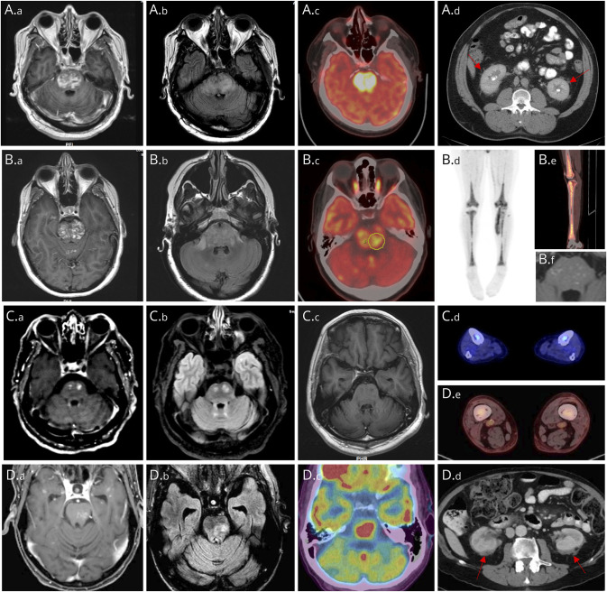
Figure. Imaging Findings in Patients With ECD and CLIPPERS-Like Enhancing Brainstem Lesions.
Imaging findings from 4 patients (A–D). MRI brain findings on T1-weighted postgadolinium administration (A.a, B.a, C.a, and D.a), T2-FLAIR (A.b, B.b, C.b, and D.b) sequences, including T1-postgadolinium follow-up scans from patient 2 (B.f) and patient 3 (C.c) demonstrating favorable response to targeted therapies. 18F-FDG PET-CT demonstrated increased uptake within the pons (patient 1 [A.c], patient 2 [B.c], and patient 4 [D.c]) and distal femur and tibia (patient 2 [B.d, B.e], patient 3 [C.d], and patient 4 [D.e]) with wispy infiltration in perinephric region producing a “hairy kidney” appearance (patient 1 [A.d] and patient 4 [D.d]).

