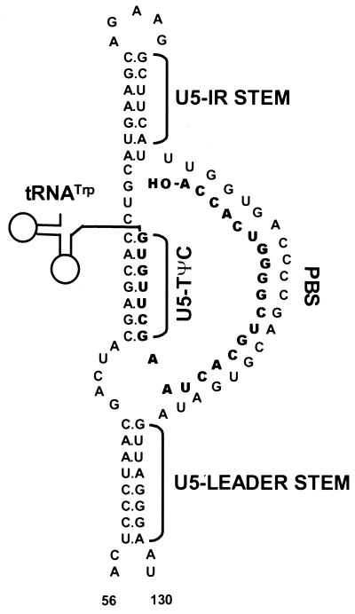
FIG. 1.
Schematic diagram of the predicted lowest-free-energy secondary structures formed when WT RSV RNA is complexed with tRNATrp (see Materials and Methods for a description of the structure prediction). Sequences are numbered from the 5′ end of the RNA genome. Only nucleotides 56 to 130 are shown, even though longer sequences were used for the predictions. tRNATrp is shown to interact with the viral RNA via two helices; one is formed between the 3′ 18 nucleotides of the tRNA and the PBS of the viral RNA, and the second is formed between the TΨC arm and loop of the tRNA and sequences in the U5 region of the viral RNA. tRNATrp sequences involved in these two interactions are shown in bold. All other tRNATrp sequences are labeled and represented by the circle-and-line drawing. The U5-IR and U5-Leader stems are also labeled.

