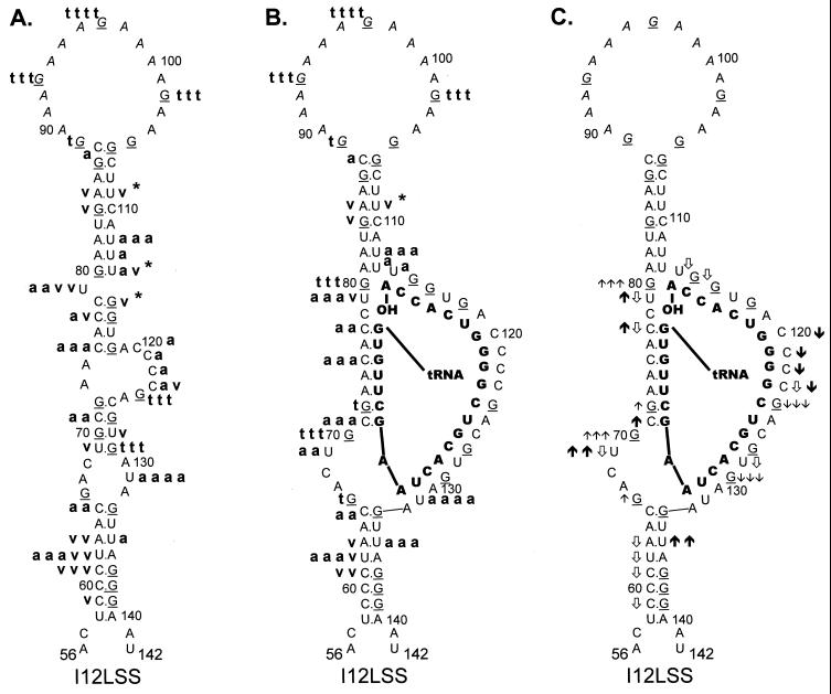
FIG. 5.
Summary of nuclease digestion data for I12Lss mutant RSV
RNA fragments. Secondary-structure diagrams for I12Lss sequences are
depicted as described in Materials and Methods. Mutant nucleotides are
denoted by italicized lettering. Other notations are as described in
the legend to Fig. 3. (A) Digestion of uncomplexed I12Lss RNA
fragments. Description of cleavages is as in the legend to Fig. 3A.
 , the exact cleavage site was
unclear, although the data did indicate that cleavage was somewhere
within the U5-IR stem. (B) Digestion of I12Lss RNA fragments complexed
with tRNATrp, where the tRNATrp sequences are
shown in bold capital letters. Description of cleavages is as in the
legend to Fig. 3B, except for V1 cleavages, where there was
no dominant cleavage with 0.0001 U of V1 per μl;
therefore, there is no v v v designation. (C) Changes in the
digestion pattern when I12Lss RNA fragments were complexed with
tRNATrp. Description of changes is as in the legend to Fig.
3C.

