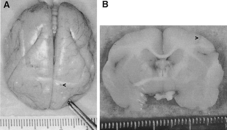
FIG. 1.
Brains from G207-injected animals. (A) Brain from T9205 at time of necropsy, 6 months after injection of 107 PFU of G207, with frontal lobes at the bottom and brain stem at the top. (B) Coronal cut through the brain from T910805 at the injection site 4 months after injection of 107 PFU of G207. In both panels, the left side of the brain is on the right side of the figure, and the injection site is marked (>). Hatch marks on rulers are in millimeters. Photographic slides were scanned with a Polaroid SprintScan 35, and Adobe Photoshop was used to adjust the contrast and brightness of the images.

