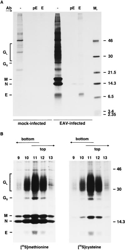
FIG. 7.
Identification of the E protein in EAV particles. (A) Analysis of pellets obtained after ultracentrifugation through a 20% (wt/wt) sucrose cushion of supernatants from mock- or EAV-infected BHK-21 cells that were labeled with 35S[Met]-35S[Cys]. Pellets were analyzed directly (−) or resuspended and subjected to immunoprecipitation analysis with E protein-specific antiserum (E) or the preimmunization serum (pE). The positions of the EAV E protein (8 kDa), N protein (apparent molecular mass, 14 kDa), M protein (16 kDa), and the small (GS) and large (GL) glycoproteins (25 kDa and 30 to 42 kDa, respectively) are shown at the left. The positions and sizes (in kilodaltons) of marker proteins (Mr) analyzed in the same gel are indicated at the right. Ab, antibody. (B) Sucrose density gradient centrifugation of [35S]Met- or [35S]Cys-labeled EAV preparations. The numbers of the gradient fractions and the position of each sample relative to the top and bottom of the centrifuge tube are indicated, as are the positions of the EAV structural proteins E, N, M, GS, and GL. Note that the N protein does not contain any Cys residues.

