Figure 1.

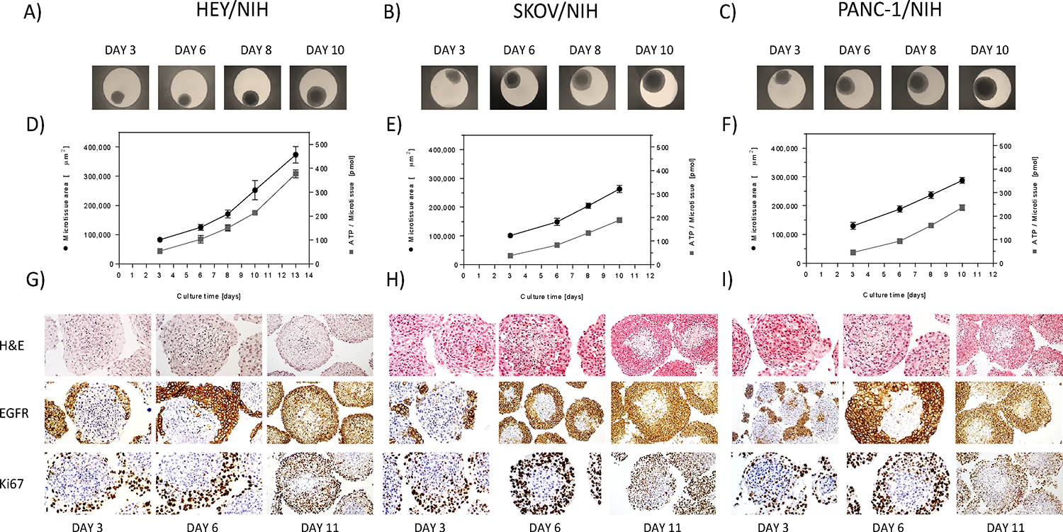
Growth profiles and morphological appearance 3D heterotypic multicellular tumor spheroids. A-C) Co-culture spheroids of HEY, SKOV-3 and PANC-1 cancer cells and NIH3T3 fibroblasts were produced with the hanging drop technology and their growth monitored over time by bright field microscopy. D-F) After 3 days of spheroid formation, tissue size (●) and intra-tissue ATP content (■) were monitored over 7 days. Each point represents the mean of 8 spheroids and their corresponding standard deviation. Bright field microscopy allowed for size assessment of the microtissues and intra-tissue ATP with CellTiterGlo™ was used as a measure of cell viability. G-I) All three heterotypic tumor microtissues were stained for histological characterization. Cancer and stromal cells were capable of reforming heterotypic, solid spheroids, as shown by hematoxylin and eosin staining. Eosin colors eosinophilic structures in various shades of red, pink and orange, whereas hematoxylin colors nuclei of cells blue. After 11 days of culture, tumor microtissues formed a necrotic area in the center of the spheres, composed of cells undergoing apoptosis and or necrosis, most likely due to hypoxia. The spheroids showed a high expression of EGRF within the cancer cells. Heterotypic tumor spheroids exhibited a spherical geometry with a central core of non-proliferating fibroblasts and proliferating cancer cells in the periphery. Differences of the individual cell types could be clearly displayed by IHC staining of Ki67.
