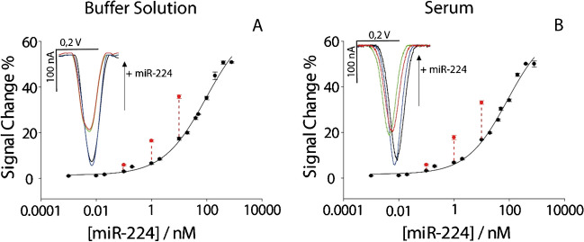
Fig. 3.
Calibration curves showcasing the effects of preconcentration on target detection. A Calibration curve in buffer solution. B Calibration curve in human serum. All the experiments have been carried out in triplicate. Insets display voltammograms illustrating target presence and concentration effects: blue curve indicates the absence of target, black curve represents the presence of a 1 nM target, red curve shows a 1 nM target preconcentrated 10 times, and green curve denotes the presence of a 10 nM target

