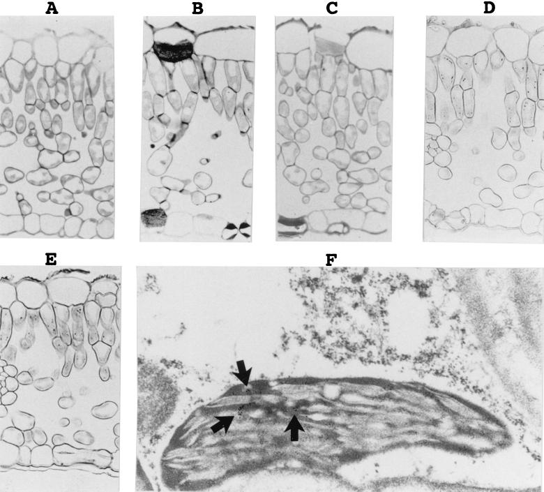
FIG. 2.
Detection of PLMVd by in situ hybridizations with DIG-labeled riboprobes. (A to E) Observations of the DIG-labeled hybrids by light microscopy. The micrographs show sections of PLMVd-infected leaves hybridized in either the absence (A) or the presence (B) of DIG-PSTVd riboprobe, a section of a healthy peach leaf probed with a minus-polarity PLMVd riboprobe (C), and sections of PLMVd-infected peach leaves probed with either the plus (D) or minus (E) PLMVd riboprobes. Control panels (A to C) were overstained to ensure the detection of trace amounts of PLMVd strands. (F) Typical electron micrograph of the hybridization of PLMVd-infected peach leaves with the minus-strand PLMVd riboprobe. The arrows point to clusters of grains representing PLMVd accumulated strands.

