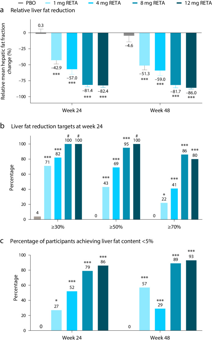
Fig. 1. Change in liver fat.

a, Relative liver fat reduction. Results are shown as LSM ± s.e.m. (n = 19 (PBO), n = 20 (1 mg RETA), n = 19 (4 mg RETA), n = 22 (8 mg RETA) and n = 18 (12 mg RETA)). b, The percentage of participants achieving liver fat reduction targets at week 24. c, The percentage of participants achieving liver fat content <5%. Comparisons versus PBO were done by using two-sided z-tests without multiplicity adjustment. *P < 0.05 versus PBO; ***P < 0.001 versus PBO; #, not calculable. Fewer participants had MRIs at week 48 (n = 8 (PBO), n = 9 (1 mg RETA), n = 9 (4 mg RETA), n = 8 (8 mg RETA) and n = 9 (12 mg RETA)) compared with week 24 (n = 14 (PBO), n = 16 (1 mg RETA), n = 15 (4 mg RETA), n = 17 (8 mg RETA) and n = 15 (12 mg RETA)).
