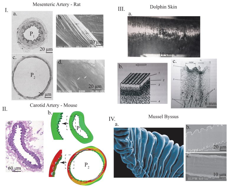
FIG. 1.
Representative images of wrinkling surfaces in various biologic systems. I. Muscular artery from rat mesentery. a. and c. show the fixed histology at low (P1) and high (P2) blood pressures while b. and d. show SEM images of silicone casts of similar non-fixed arteries at equivalent pressures to the histology slides. In both sets of images, it is apparent that wrinkle and fold patterns appear on the inside of the artery at lower pressures and disappear as the artery distends with higher pressure (adapted with permission from [11]). II a. Histology of mouse carotid artery with luminal (inner) wrinkling. b. and c. show numerical simulations where this exact artery geometry is pressurized, showing the smoothing out of luminal wrinkles with increasing luminal pressure, showing the generality of the mechanism. III. Dolphin skin with periodic macroscopic wrinkles (a.) and the representative histology (b.) and (c.), adapted with permission from [13]. IV. Mussel byssus showing the intricate external wrinkles with SEM (a.) and the two states of the byssus under different conditions wrinkles (b.) and flat (c.), adapted from [5].

