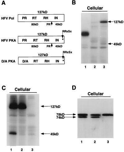
FIG. 1.
IP-PKA and Western blot analysis of HFV-PKA and HFV-D/A-PKA. Assay conditions are as described in Materials and Methods except that the second IP step was omitted for PKA analysis of cellular Pol proteins. Anti-RH antiserum was used to immunoprecipitate Pol, and anticapsid antiserum was used for Gag Western blots. (A) Schematic diagram of HFV (wt) and HFV-PKA Pol proteins. (B) IP-PKA analysis of HFV-PKA. Lanes: 1, positive control for PKA phosphorylation; purified interleukin-1 receptor (38); 2, HFV (wt)-transfected cells; 3, HFV-PKA-transfected cells. (C) PKA analysis of cell lysates mock transfected (lane 3) or transfected with HFV-PKA and HFV-D/A-PKA (lanes 1 and 2, respectively). (D) Western blot analysis of HFV-PKA Gag proteins. Lanes: 1, HFV (wt); 2, HFV-PKA; 3, HFV-D/A-PKA.

