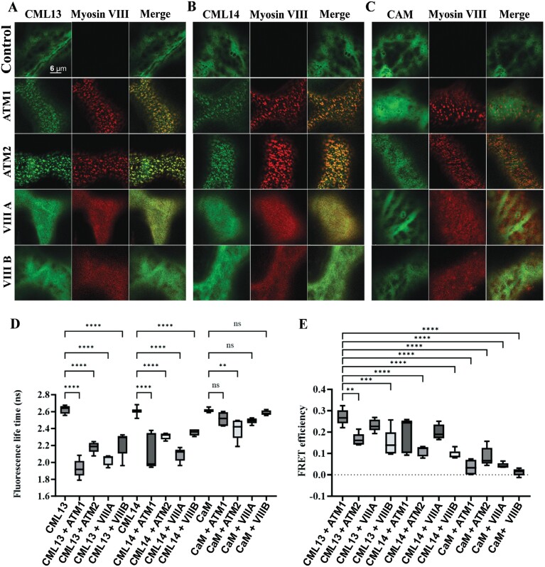
Fig. 3.
FRET-FLIM analysis of myosin VIII IQ-tail fragments with CML13, CML14, and CAM. RFP–ATM1IQ-tail, RFP–ATM2 IQ-tail, RFP–VIII-A IQ-tail, or RFP–VIIIB IQ-tail were transiently expressed in N. benthamiana leaves with (A) GFP–CML13, (B) GFP–CML14, or (C) GFP–CaM. (D) Fluorescence lifetime. (E) FRET efficiency. Microscopy was performed 48 h after agro-infiltration using a Leica SP8 confocal microscope or Leica Stellaris 8 with a white laser and Falcon application. Statistical analysis was by one-way ANOVA, **P<0.01, *** P<0.001, ****P<0.0001. All microscopy images are at the same magnification as indicated in panel A.

