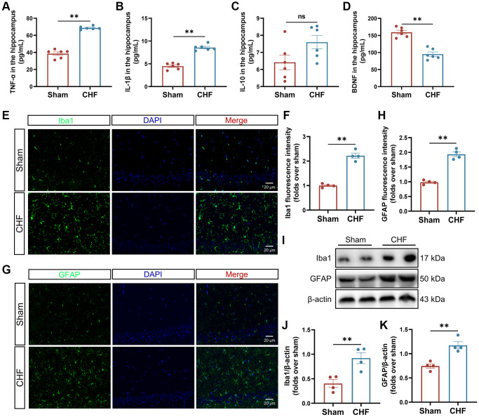
Figure 3.
CHF triggers the inflammatory responses via the activation of microglia and astrocytes. (A–D) The content of TNF-α (A), IL-1β (B), IL-10 (C), and BDNF (D) in the hippocampus was detected by ELISA n = 6. (E, F) Iba1 immunofluorescence staining in the hippocampus and their quantitative analysis. Green: Iba1; Blue: DAPI. Scale bars: 20 μm n = 4. (G, H) GFAP immunofluorescence staining in the hippocampus and their quantitative analysis. Green: GFAP; Blue: DAPI. Scale bars: 20 μm n = 4. (I–K) Representative Western blots (I) and quantification data of Iba1 (J) and GFAP (K). β-actin was used as a loading control. Data are presented as the Mean ± SEM; *P < 0.05, and **P < 0.01 vs. Sham; n = 3.

