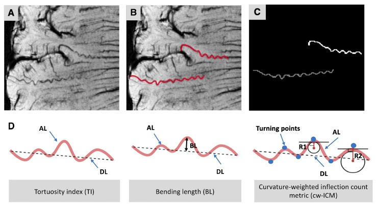
Figure 1.
Quantitative tortuosity measurements of the medullary artery. (A) Post-contrast susceptibility-weighted imaging (SWI) data with minimum intensity projection of four (mIP = 4) clearly showed tortuous cerebral small arteries. (B) Two different tortuous small arteries were manually segmented assisted with ITK-SNAP toolbox (red). (C) Binary masks of the two vessels of interest (VOIs) were acquired for further centerline tracking and morphological analyses. (D) Quantitative morphological measurements include tortuosity index (TI), bending length (BL), and curvature-weighted inflection count metric (cw-ICM). TI = actual length (AL) / direct length (DL); BL is the maximum perpendicular distance between AL and DL; cw-ICM = maximum curvature (i.e., ) × turning points × TI.

