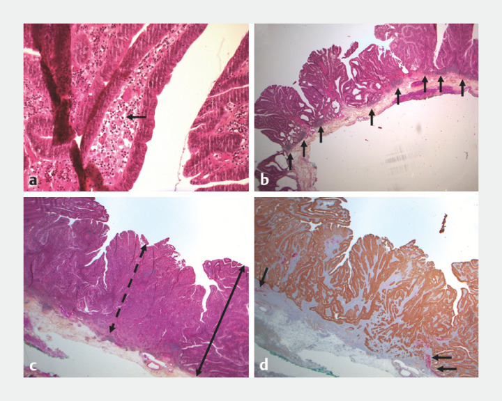
Fig. 5.
Microscopic examination of the resection specimen containing a, b chicken skin and c,d green sign. Macrophagic infiltration with xanthomatous morphology (black arrow in a ), as previously described in other studies. Increased number of lymphoid nodules at the periphery of invasive carcinoma (black arrow in b ) corresponding to a hyperplastic reaction of the gut associated lymphoid tissue, which could at least partially explain the chicken skin with regularly scattered small nodules lifting the mucosa. Thinning of the mucosa (dotted double arrow in c ) compared with the adenomatous mucosa (double arrow in c ) as invasive glands destroy the mucosa. Destruction of the muscularis mucosae (black arrow in d , in red) by invasive glands which may contribute to the increase in the hemoglobin detection signal, resulting in the green sign.

