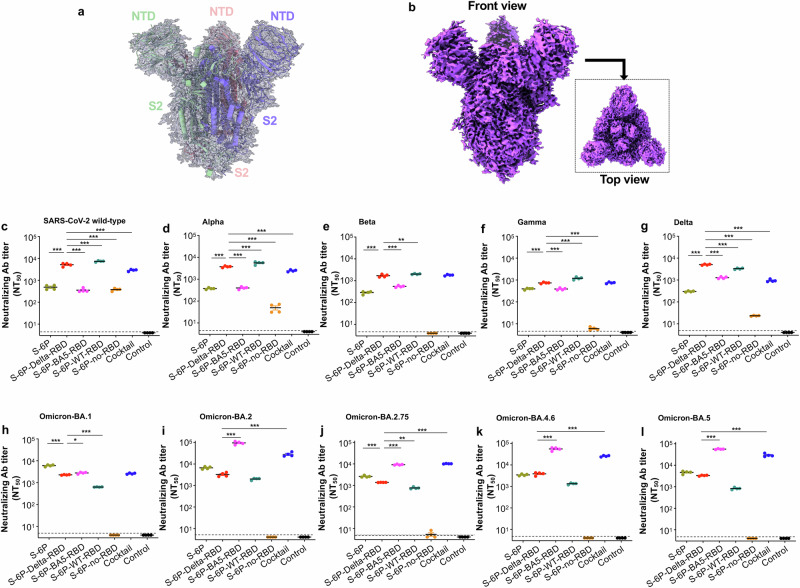
Fig. 2. Cyro-EM structures of S-6P-no-RBD protein and neutralizing ability of subunit vaccines.
a Overview of the cryo-EM map of the truncated spike (S) (i.e., S-6P-no-RBD) protein with structural model inside. The structure is presented in cartoons with tube helices. b Front and top views of the cryo-EM map. c–i Neutralizing antibodies induced by subunit vaccines against multiple variants and ancestral strain of SARS-CoV-2. The purified proteins, including S-6P, S-6P-Delta-RBD, S-6P-BA5-RBD, S-6P-WT-RBD, and S-6P-no-RBD (10 μg/mouse), or PBS control, were intramuscularly (i.m.) injected into K18-hACE2 mice in the presence of adjuvants. The cocktail consisted of the S-6P-Delta-RBD and S-6P-BA5-RBD proteins (5 μg/protein; 10 μg/mouse) with the adjuvants. The mice were boosted twice at 3-week intervals with the same immunogen and adjuvants, as described in Fig. 1. Sera collected from 10 days after the 3rd immunization were evaluated for neutralizing antibodies (Abs) against pseudoviruses encoding the S protein of the ancestral (wild-type, WT) SARS-CoV-2 strain (c), Alpha (d), Beta (e), Gamma (f), and Delta (g) variants, as well as the Omicron BA.1 (h), BA.2 (i), BA.2.75 (j), BA.4.6 (k), and BA.5 (l) subvariants. The NT50 is expressed as 50% neutralizing Ab titers against each pseudovirus infection in 293T cells expressing human angiotensin converting enzyme-2 (hACE2/293T). The data are presented as the mean ± standard deviation of the mean (s.e.m.) of four wells from pooled sera of five mice in each group. The limit of detection for the neutralization assay was 1:5 (c–l). Ordinary one-way ANOVA (Dunnett’s multiple comparison test) was used to compare the statistical differences of neutralizing antibody titers induced by S-6P-Delta-RBD and other vaccination groups. *P < 0.05, **P < 0.01, and ***P < 0.001 designate significant differences between S-6P-Delta-RBD and other vaccination groups. The experiments were repeated twice (c–e, h) or three times (f, g, i–l) to confirm the results.

