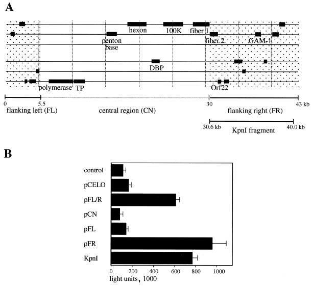
FIG. 1.
E2F activation assay with different CELO virus constructs. (A) Schematic organization of the CELO virus genome. Some of the conserved E2 and late genes are shown. The shaded regions flanking the central part (CN) on the left and right (FL and FR) indicate sequences that are unique to CELO virus. (B) Reporter construct E2-Luc (0.2 μg) was transfected into CEF cells as described in Materials and Methods (control lane). For cotransfection assays, 0.4 μg of the following plasmids were transfected in addition to E2-Luc: pCELO (full-length CELO virus sequence), pFL/R (kb 0 to 5.5) and 30 to 43 of CELO sequence), pCN (kb 5.5 to 30 of CELO sequence), pFL (kb 0 to 5.5 of CELO sequence), pFR (kb 30 to 43 of CELO sequence), and KpnI (kb 30.6 to 40.0 of CELO sequence); 24 h after transfection, 20 μl of cell lysate was analyzed for luciferase activity.

