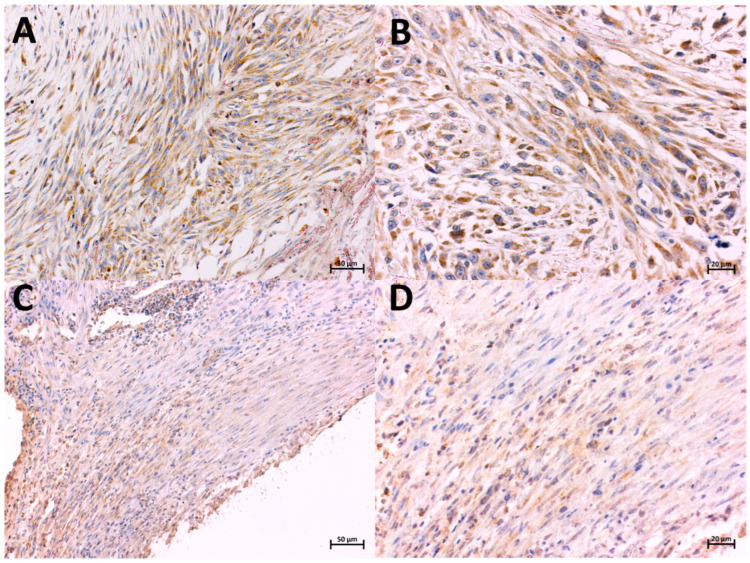
Figure 1.
Localization of COX-2-expressing cells in FISS (A,B) and non-FISS (C,D), immunohistochemically stained. Brown color indicates positive staining. Positive immunostaining for COX-2 in FISS, intensity 3, 90% of positive cells, magnification 20× and 40×, respectively (A,B). Positive immunostaining for COX-2 in non-FISS, intensity 1, 60% of positive cells, magnification 20× and 40×, respectively (C,D).

