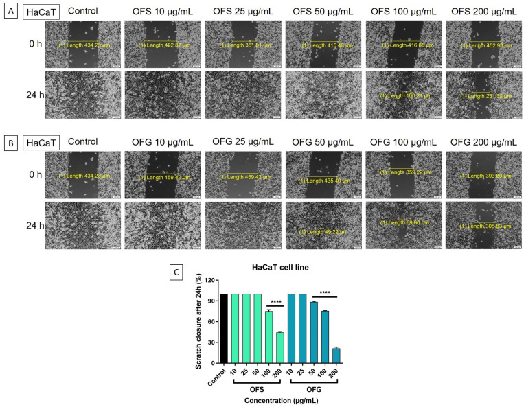
Figure 11.
HaCaT keratinocytes—microscopic images of the cells treated with the OF extracts (10, 25, 50, 100, and 200 μg/mL), initially at 0 h and 24 h; pictures were taken by light microscopy at 10x magnification. (A) Cells treated with OFS extract; (B) cells treated with OFG extract; (C) graphical representation of the scratch closure rate at 24 h post-treatment with OF extracts. Results are expressed as mean ± SD. Comparison among groups was made using one-way ANOVA and Dunnett’s multiple comparison post-test (**** p < 0.0001 vs. control).

