Table 1.
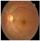
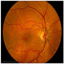
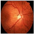
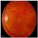
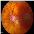
Tabular depiction of AMD fundus image stages showcasing a comparison along with visual samples.
| No AMD (Normal) | Early | Intermediate AMD | Advanced Dry AMD | Advanced Wet AMD |
|---|---|---|---|---|
| Refers to images showing no signs of AMD-related changes, often depicting individuals without clinical evidence of AMD or those in early stages of the disease with minimal abnormalities, such as a small number (5–15) of small (<63 m) or absent Drusen without pigment alterations. | Early AMD clinically presents with Drusen and abnormalities in the retinal pigment epithelium (RPE). It may involve one or both eyes, showing pigmentary changes, a few small Drusen, and/or intermediate-sized (63–124 m) Drusen. | Intermediate AMD lies between early and advanced stages, characterized by pigmentary changes and Drusen, small yellow deposits beneath the retina. It may include one large druse (>125 m), one extensive intermediate-sized druse (20 soft or 65 hard without any soft), and/or GA excluding the macula in one or both eyes. | GA is marked by the gradual loss of RPE cells in specific macular areas, forming well-defined atrophic patches. It results in progressive central vision loss and is a major cause of significant visual impairment in AMD patients. | Wet AMD is an advanced and severe form of the disease, characterized by the growth of abnormal blood vessels beneath the retina. These vessels can leak fluid or blood, causing retinal damage and rapid vision loss if untreated. It commonly results in sudden and severe central vision impairment. |
|
|
|
|
|
