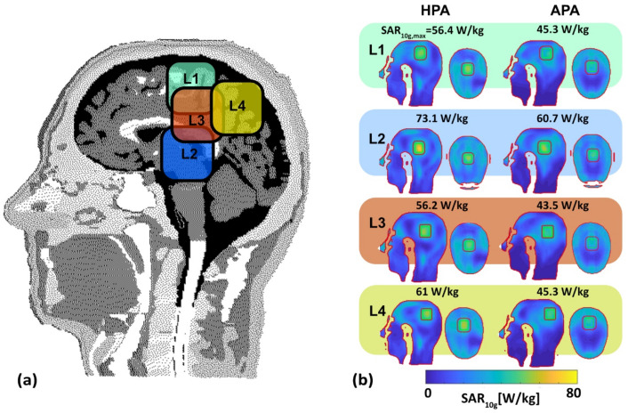
Figure 6.
Evaluation of SAR distribution in the human head voxel model Duke. (a) Definition of the locations L1–4 of the target regions (TR) placed in the human head voxel model Duke highlighted in four different colors, (b) SAR2q maps obtained for the HPA and the APA for the four TR locations (green borderline, volume: 37.5 mm × 37.5 mm × 4 mm = 5.625 cm3) placed in the human head voxel model Duke employing MVFS while integrating the total exposure from two to three excitation configurations, which will be executed in a sequentially alternating pattern over time for maximization of SAR10g inside the TR. The size of the safe margin (red borderline) between target and healthy tissues was set to 10 mm. SAR10g,max inside the TR is annotated for the four TR locations for the HPA and APA RF applicator.

