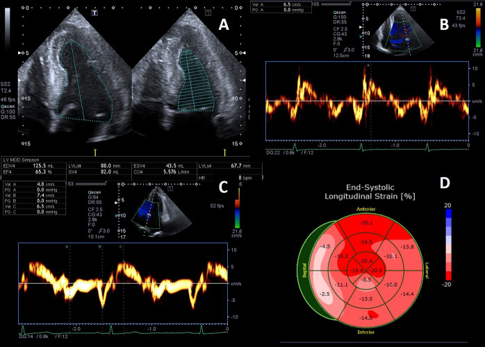
Fig. 3.
Example of Fabry cardiomyopathy with normal LV ejection fraction (LVEF) but impaired indexes of longitudinal systolic function. (A) Apical four chambers view: LVEF 65% measured by biplane Simpson method; (B, C) Tissue Doppler mitral annular velocities at lateral and septal corners respectively, showing low systolic velocities (6.5 cm/s at both sides); (D) 2D speckle tracking analysis bull’s-eye plot, showing reduced LV-GLS value (–15%).

