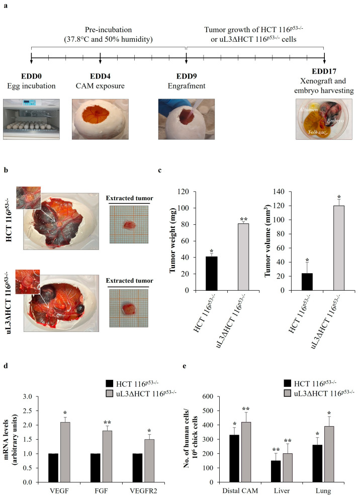
Figure 1.
Angiogenic and metastatic potential of uL3∆HCT 116p53−/− cells in Chorioallantoic Membrane (CAM) model. (a) General workflow of in ovo CAM assay. Fertilized eggs were incubated at 37.8 °C. Eggs were opened on egg development day (EDD) four (EDD4). After 5 days, HCT 116p53−/− or uL3∆HCT 116p53−/− cells in Matrigel pellets (3 × 10⁶ tumor cells per pellet) were applied to the CAM, and xenografts were allowed to develop and grow for 8 days. On EDD17, CAM xenografts were harvested, embryos were sacrificed, and chick embryo tissues and xenografts were collected for further processing. (b) Representative macroscopic images of HCT 116p53−/− and uL3∆HCT 116p53−/− cell line-derived tumors on EDD17. (c) Quantification of tumor weight and tumor volume of HCT 116p53−/− and uL3∆HCT 116p53−/− cell-derived tumors is shown. * p < 0.05; ** p < 0.01. (d) Expression of VEGF, FGF and VEGFR2 in the proximal CAM of HCT 116p53−/− and uL3ΔHCT 116p53−/− xenografts was quantified by RT-qPCR using specific primers (Table 1). Bars represent the mean of triplicate experiments; error bars represent the standard deviation. * p < 0.05; ** p < 0.01. (e) qPCR quantification of human HCT 116p53−/− and uL3ΔHCT 116p53−/− cells in the distal CAM, liver and lung tissues using universal human Alu (h-Alu) primers. As internal control used to validate the presence of an equivalent amount of chick genomic DNA (gDNA), we employed chick GAPDH (ch-GAPDH) primers (Table 2). Bars represent the mean of triplicate experiments; error bars represent the standard deviation. * p < 0.05; ** p < 0.01.

