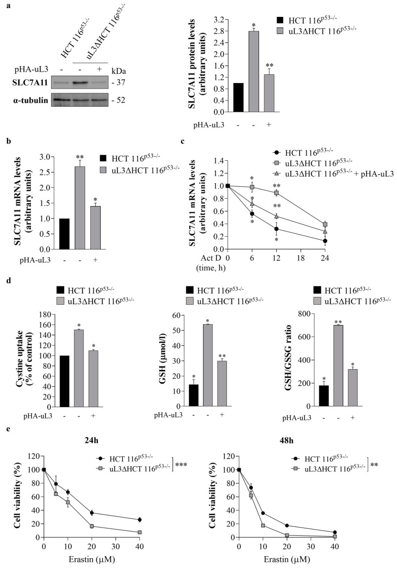
Figure 4.
Effect of uL3 silencing on SLC7A11 expression and erastin cytotoxic activity. (a) Protein extracts from HCT 116p53−/− cells and uL3∆HCT 116p53−/− cells transfected or not with pHA-uL3 were analyzed by western blotting (WB) with anti-SLC7A11 antibody; α-tubulin was used as loading control. Full-length blots are shown in Figure S7. The graph shows the relative densitometric analyses, expressed as arbitrary units. Bars represent the mean of triplicate experiments; error bars represent the standard deviation. * p < 0.05, ** p < 0.01 vs. HCT 116p53−/− cells set at 1. (b) RT-qPCR of total RNA extracted from HCT 116p53−/− cells and uL3∆HCT 116p53−/− cells transfected or not with pHA-uL3 using specific primers for SLC7A11 (Table 1). * p < 0.05, ** p < 0.01 vs. HCT 116p53−/− cells set at 1. (c) HCT 116p53−/− cells and uL3∆HCT 116p53−/− cells transfected or not with pHA-uL3 were treated with actinomycin D (Act D) (5 μg/mL). At the indicated time points (0, 6, 12 and 24 h), total RNA was isolated, and mRNA levels of SLC7A11 and β-actin were determined by RT-qPCR by using specific primers (Table 1). The relative amount of SLC7A11 mRNA in untreated cells was set to 1, and the levels of SLC7A11 mRNA in cells treated with Act D were calculated accordingly. * p < 0.05, ** p < 0.01 vs. untreated cells. (d) Evaluation of cystine uptake, reduced glutathione (GSH) levels and GSH/ oxidized glutathione (GSSG) ratio in HCT 116p53−/− cells and uL3∆HCT 116p53−/− cells transfected or not with pHA-uL3 for 24 h. * p < 0.05, ** p < 0.01 vs. HCT 116p53−/− cells. (e) HCT 116p53−/− and uL3∆HCT 116p53−/− cells were treated or not with different concentration of erastin (from 5 to 40 µM) for 24 and 48 h. After incubation, cell viability was evaluated using the MTT assay. Cell viability of untreated cells was set to 100%. Bars represent the mean of triplicate experiments; error bars represent the standard deviation. ** p < 0.01, *** p < 0.001 uL3ΔHCT 116p53−/− cells vs. HCT 116p53−/− cells.

