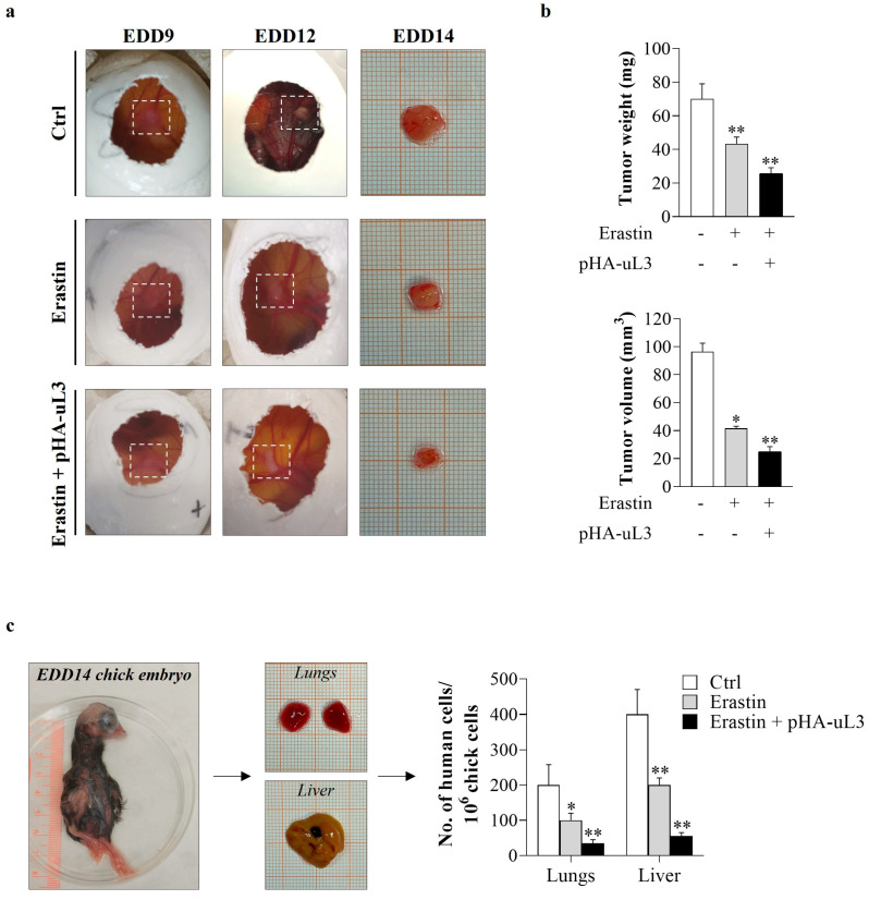
Figure 7.
Antitumor effects of erastin plus uL3 in CAM model. uL3∆HCT 116p53−/− cells, untransfected or transfected with pHA-uL3, were engrafted onto CAM surface on EDD9 and then treated or not with erastin (10 µM). (a) Macroscopic images of uL3∆HCT 116p53−/− cell line-derived tumors were captured on EDD9, 12 and 14. Selected areas of CAM in which uL3∆HCT 116p53−/− cells were engrafted are shown. (b) On EDD14, excised tumors were weighed and measured using a digital caliper in order to determine the effect of the treatments on tumor growth. * p < 0.05, ** p < 0.01 vs. untreated cells-derived tumors. (c) Representative images of EDD14 chick embryo and dissected lungs and liver. gDNA was extracted and analyzed by qPCR using specific primers for h-Alu. As internal control used to validate the presence of equivalent amount of chick gDNA, we employed ch-GAPDH primers (Table 2). * p < 0.05, ** p < 0.01 vs. control cells.

