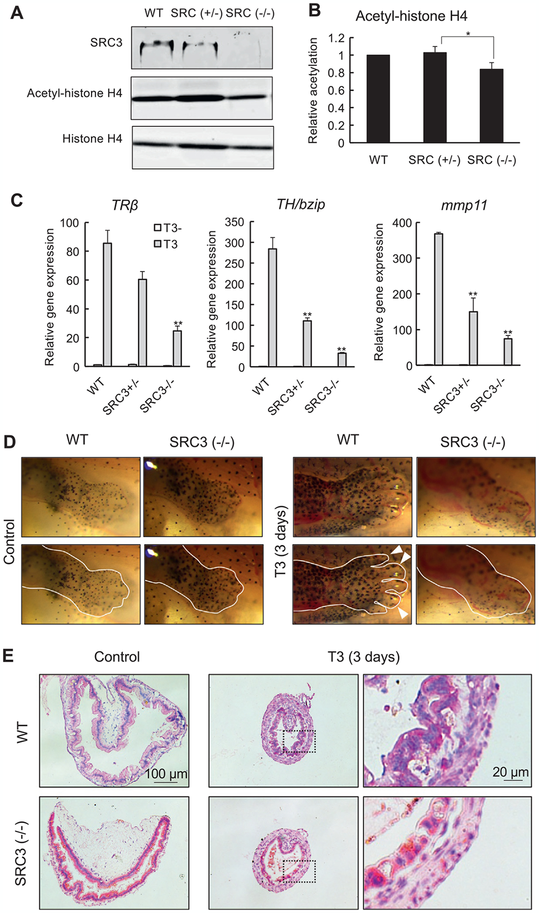
Fig. 4.

SRC3 knockout reduces histone H4 acetylation and inhibits T3-induced metamorphosis. (A)/(B) Western blots showing that homozygous SRC3 knockout tadpoles have no detectable SRC3 protein and reduced histone H4 acetylation. Total protein was isolated from whole body of wild type (WT), heterozygous (SRC3+/−), and compound heterozygous (functionally homozygous, SRC3−/−) SRC3 mutant tadpoles at stage 46 and subjected to Western blot analyses with antibodies against SRC3, acetyl-histone H4, or histone H4 (A). The signal intensity of the bands was measured by image j and presented as the mean ± standard deviation of n = 3 (B). Statistical analysis: ANOVA one-way followed by Tukey analysis. *P <0.05. (C) SRC3 knockout reduces gene activation by T3 in the intestine. The intestine was isolated from wild type (WT), SRC3+/− and SRC3−/− tadpoles at stage 54 with or without 18 h T3 treatment before RNA isolation and qRT-PCR analysis of the indicated TR target genes. **P < 0.01 for T3 treated vs. WT intestine. (D) SRC knockout inhibits T3-induced limb metamorphosis. Wild type (WT) and SRC3 knockout (SRC3−/−) tadpoles at stage 54 were treated with 10 nM T3 for 3 days at 25°C and hindlimb region was photographed. The lower panels were the same as the top panels except the added white line marking the approximate boundary of the outer limb epithelium and/or the boundaries of the digits (white arrowheads indicate some of the digits of the wild type animal treated with T3). (E) Adult intestinal epithelium development is inhibited by SRC3 knockout. Cross-sections of the intestine in WT and SRC3−/− tadpoles as in (D) were stained with methyl green-pyronin Y (staining DNA blue and RNA red). Note that the adult epithelium folds began to form after 3 days of T3 treatment in WT but not SRC3−/− tadpoles. See Tanizaki, Bao, et al. (2021) for more details.
