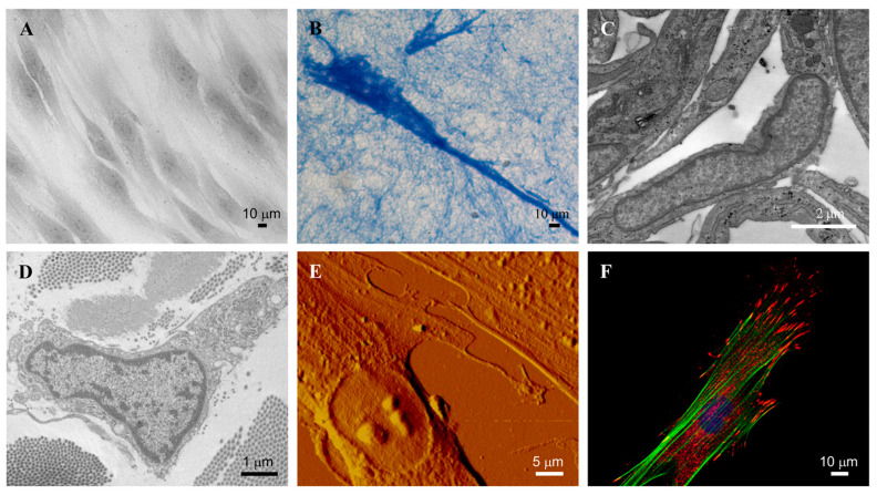
Figure 1.
Representative images of dermal fibroblasts. Fibroblasts in 2D (A) and 3D (B) culture observed by light microscopy. Fibroblast in culture (C) and in healthy skin (D), observed by transmission electron microscopy. Fibroblast in culture observed by atomic force microscopy (E) and by confocal microscopy (F). Images are from authors’ laboratory.

