Figure 1.
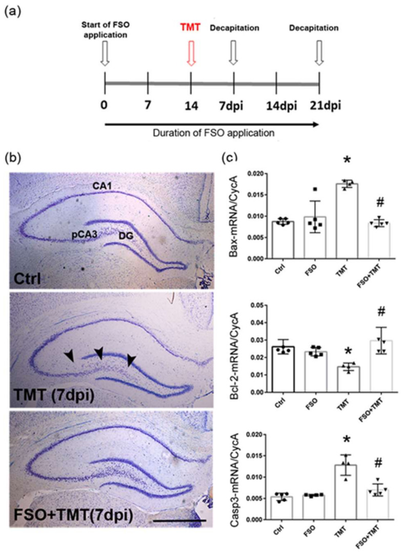
FSO prevented TMT-induced hippocampal damage 7 days post intoxication. (a) A schematic illustration of an experimental design. (b) Representative thionin-stained coronal sections obtained from control (Ctrl), trimethyltin (TMT), and flaxseed oil (FSO)-treated TMT-intoxicated (FSO+TMT) animals. Arrowheads indicate injured neuronal cell layers in the hippocampus, while the laminar organization of pCA3 subfield was regular in the FSO-treated TMT sections. Scale bar = 500 μm. (c) The abundance of transcript coding Bax, Bcl-2, and Caspase3 (Casp3) assessed by RT-qPCR in Ctrl, FSO, TMT, FSO+TMT animals. Bars represent mean target mRNA abundance relative to CycA (± SD) from n = 5 hippocampi per group. Significance shown inside the graphs: * p < 0.05 or less relative to Ctrl; # p < 0.05 or less relative to TMT. pCA3, proximal CA3 subfield of the hippocampus; DG, dentate gyrus; dpi, days post intoxication.

