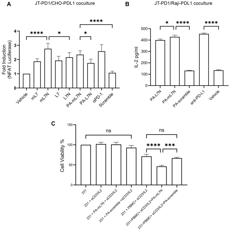
Figure 4.
Evaluation of the efficacy of L7 and L7 analogues in reinvigorating PD-1-exhausted T cells and promoting cancer cell killing by PBMCs. (A) The indicated peptides (10 μM) were added to the JT-PD1/CHO-PDL1 coculture, followed by NFAT luciferase assays to determine T-cell activation. A PD-L1 antibody was included for comparison. (B) The PA-tagged versions of both L7N and mL7N significantly reinvigorated T cells in the JT-PD1/Raji-PDL1 coculture. Shown are IL-2 levels 24 h post addition of the peptides or antibody. (C) PA-mL7N significantly promoted killing of MDA-MB-231 cells by PBMCs ex vivo while exhibiting no apparent toxicity to the cancer cells at 10 μM. Shown are cell viability data of MDA-MB-231 cells under the indicated conditions measured by WST-8 assay. *, p < 0.01; ***, p < 0.0001; ****, p < 0.00001; one-way ANOVA test.

