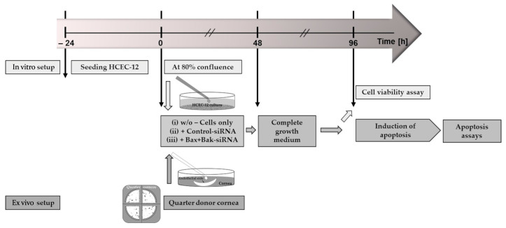
Figure 2.
Schematic representation of the time course of the experiments. For the in vitro approach, corneal endothelial cells (HCECs) were seeded for 24 h at a density of 2 × 10⁵ to reach 80% confluence for the experiment. For the ex vivo approach, the donor cornea was quartered, and one quarter was placed with the endothelial side up in a well (“cornea in a cup”). The procedure for both cultures (highlighted in green) was then to apply siRNA in serum-free medium at time 0 h with the following treatment: (i) no addition of siRNA as control (Cells only); (ii) addition of non-target siRNA (Control-siRNA); or addition of siRNA to knockdown the pro-apoptotic proteins Bax and Bak (Bax+Bak-siRNA). After 48 h, the medium in both cultures was changed, and the appropriate complete growth medium was added. After a further 48 h of knockdown, experiments were performed for the HCEC viability assay. In order to analyze the transfection, cell death was induced with etoposides (HCECs additional with staurosporine), followed by apoptosis assays such as TUNEL.

