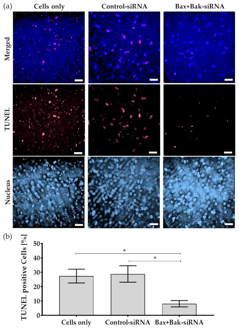
Figure 8.
As a proof-of-principle, apoptosis was detected by TUNEL (TdT-mediated dUTP-biotin nick end labeling) assay in the endothelium of donor corneas after transfection with Accell Control-siRNA or Bax+Bak-siRNA compared to untransfected cells (Cells only). After incubation for 96 h, the apoptosis inducer etoposide (42.5 µM for 21 h) was added. (a) Exemplary images of the nuclei of the cells labeled with TUNEL (red) and Dapi (blue). Note that despite the induction of apoptosis, fewer nuclei of apoptotic cells were visible in the corneal endothelium compared to controls—Cells only and Control-siRNA. (LSM780, Zeiss; red: TUNEL—apoptotic DNA fragmentation; blue: Dapi—cell nuclei; 40× objective, zoom 0.6, 3D overlay, bar = 40 µm). (b) Quantification indicated that the apoptosis rate was significantly reduced in corneas after the Bax+Bak knockdown. (ImageJ; n = 3, mean ± s.e.m., RM one-way ANOVA posthoc uncorrected Fisher’s LSD test: * p < 0.05).

