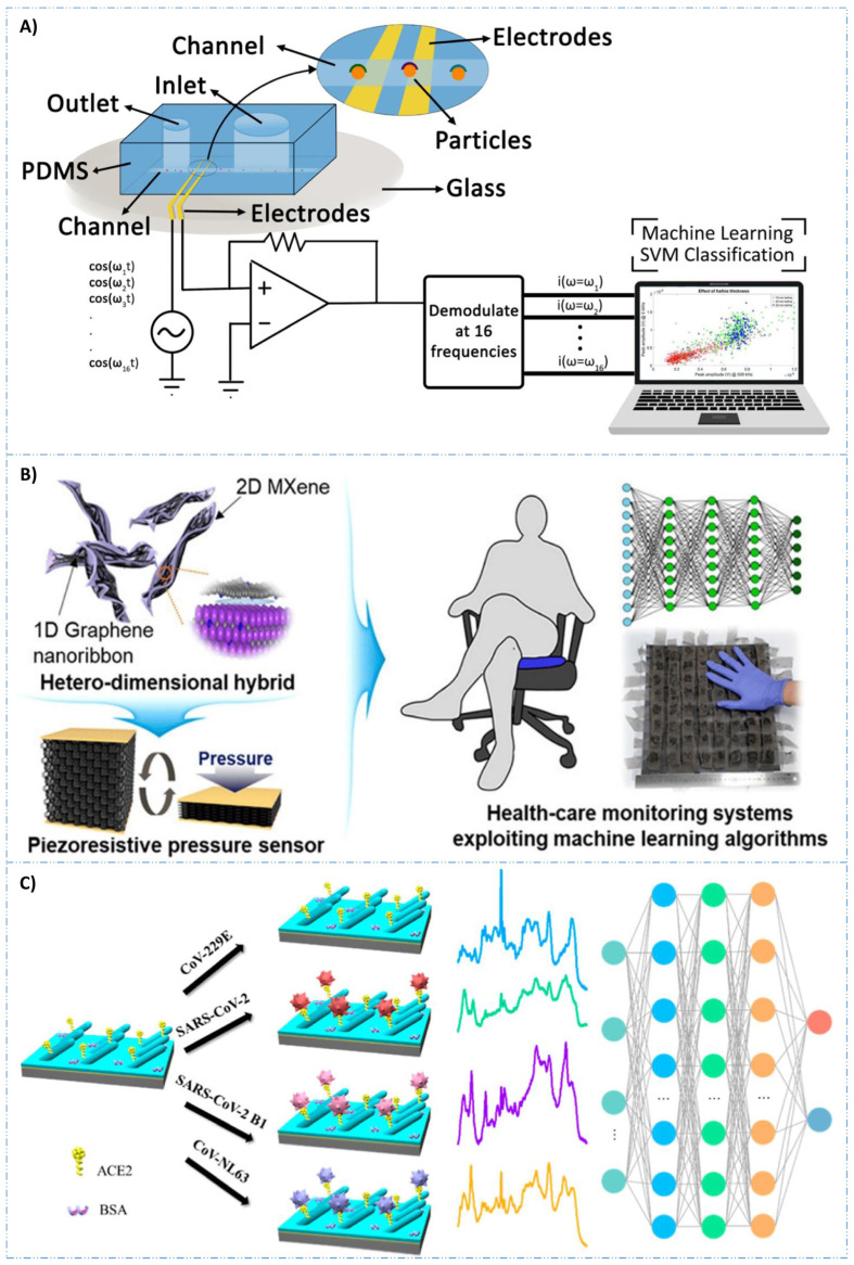
Figure 5.
(A) Scheme of electrical impedance cytometer. As cells pass from the inlet to the outlet in these biosensors, alterations in impedance are detected by a lock-in amplifier. This amplifier can simultaneously apply signals at various frequencies. Subsequently, the data are recorded and analyzed using SVM. Reproduced with permission from [155]. (B) Interfacing 1D graphene nanoribbons with 2D MXene for the development of pressure biosensor, trained using ML algorithm. Reproduced with permission from [157]. (C) Schematic illustration of angiotensin converting enzyme 2 (ACE2)-functionalized AgNR@SiO2 array for SARS-CoV-2 variant detection. Reproduced with permission from [162].

