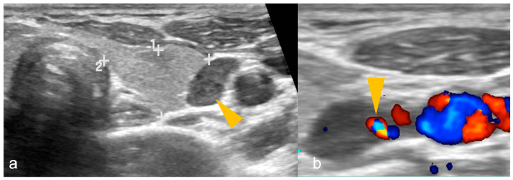
Figure 1.
(a,b): Parathyroid adenoma on ultrasonography in a patient with primary hyperparathyroidism. (a) A well-defined oval-shaped homogeneously hypoechoic lesion (arrowhead) lateral to the left lobe of thyroid gland (shown by 1, 2 and + sign). (b) Colour Doppler image shows feeding vessel sign (arrowhead). Imaging findings are suggestive of parathyroid adenoma.

