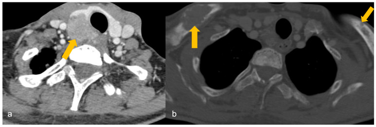
Figure 5.
A 32-year-old male with a family history of parathyroid carcinoma presented with elevated serum calcium and parathyroid hormone. (a) Heterogeneously enhancing mass (arrow) arising posterior to the right lobe of thyroid gland on contrast-enhanced computed tomography (CECT), infiltrating the thyroid lobe and occupying the right trachea-oesophageal groove, findings suggestive of parathyroid carcinoma. (b) Osteolytic lesions in bilateral clavicles (arrows) on CECT, suggestive of biopsy-proven brown tumours.

