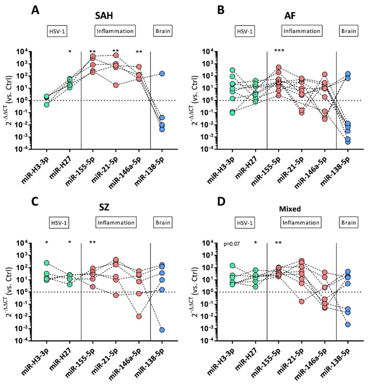
Figure 4.
miRNA expression profiles of CSF-derived exosomes’ preparations. Log10 fold changes of HSV-1-derived miR-H3-3p and miR-H27 (green symbols), cellular inflammatory miR-155-5p, miR-21-5p, miR-146a-5p (red symbols), and brain-derived miR-138-5p (blue symbols) are shown as dot plots in SAH (A), AF (B), SZ (C), and non-traumatic/non-psychiatric patients (D). Log10 fold changes were calculated using the 2−ΔΔCT method. Control (Ctrl)-derived exosomal preparations served as reference, and the dotted horizontal line (fold change = 1) represents the cut-off for elevated transcripts. For viral miR-H3-3p, n = 5 SAH, n = 10 AF, n = 5 SZ, and n = 7 non-traumatic/non-psychiatric patients were analyzed and normalized against n = 3 Ctrl samples. For viral miR-H27, n = 5 SAH, n = 7 AF, n = 5 SZ, and n = 7 non-traumatic/non-psychiatric patients were analyzed and normalized against n = 4 Ctrl samples. For host miRNAs, n = 5 SAH, n = 11 AF, n = 6 SZ, and n = 8 non-traumatic/non-psychiatric patients were analyzed and normalized against n = 5 Ctrl samples. Wilcoxon rank-sum test was performed to analyze differences against controls (Ctrl). Significant differences are marked by * p ≤ 0.05, ** p ≤ 0.01, and *** p ≤ 0.001.

