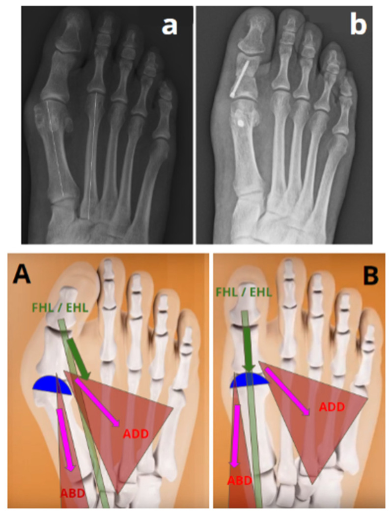
Figure 3.
Representation of a spontaneous reduction in the IMA after 3D chevron osteotomy without lateral translation of the first metatarsal head (“successful without translation” group). ADD = adductor hallucis muscle; ABD = abductor hallucis muscle; FHL = tendon of the flexor hallucis longus muscle; EHL = tendon of the extensor hallucis longus muscle. (A) The deformity in the valgus and pronation of the distal epiphysis of the first metatarsal (represented in blue) induces an imbalance in the adjacent musculotendinous structures. (a) Preoperative radiography of a hallux valgus in the “successful correction without translation” group with an IMA at 13°. (B) An osteotomy combining supination and varization allows for a correction of this deformity, resulting in the balance of the adjacent musculotendinous structures, allowing for the spontaneous reduction in the IMA. (b) Postoperative radiography showing a reduction in the IMA at 6° without translation.

