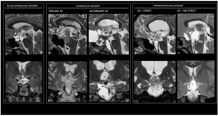
Figure 3.
Topographical categories of craniopharyngiomas. Preoperative sagittal (a–e) and coronal (f–j) T2 magnetic resonance images show the five major topographical categories of craniopharyngiomas. Sellar–suprasellar category. Sellar–suprasellar CP (a–f): the chiasmatic cistern is occupied by the tumor, the PS is not visible, the chiasm is stretched upward (thick white arrow in (a)), the mammillary body angle (MBA) is acute (65°), and the hypothalamus (thin white arrows in (f)) is around the upper-third level of the tumor; the 3 V is free of tumor. Suprasellar category. Pseudo-third ventricle CP (b,g): the sella, chiasm cistern, and 3 V are occupied by a giant tumor; the PS is not visible, the chiasm is stretched upward (thick white arrow in (b)), the mammillary body angle is obtuse (120°), and the hypothalamus (thin white arrows in (g)) is around the upper third of the tumor. Secondary intraventricular CP (c,h): the chiasm cistern and the 3 V are occupied by the tumor, the PS is not visible, the chiasm is stretched forward (thick white arrow in (c)), the angle of the mammillary body is acute (40°), and the hypothalamus is located around the mid-third of the tumor (thin white arrows in (h)). Intraventricular category. Strictly intraventricular CPs (d,i): the chiasm cistern is tumor-free, the PS is entirely visible, the chiasm is compressed downward (thick white arrow in (d)), the angle of the mammillary body is acute (35°), and the hypothalamus is in the lower-middle third of the tumor (thin white arrows in (h)). Not-strictly intraventricular CPs (e,j): the chiasm cistern is occupied by tumor, the chiasm is compressed forward (thick white arrow in e), the angle of the mammillary body is hyperacute (30°), and the hypothalamus is located around the mid-third of the tumor (thin white arrows in (j)). 3 V, third ventricle; Ch, chiasm; MBA, mammillary body angle; t, tumor.

