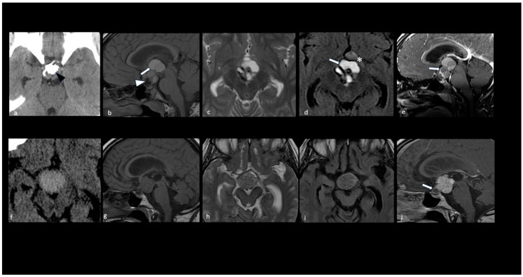
Figure 4.
Key morphologic features in adamantinomatous and papillary craniopharyngiomas. CT scan (a,f); sagittal T1 (b,g), axial T2 (c,h), axial FLAIR (d,i), and sagittal T1 with contrast medium (e,j) MRI. Adamantinomatous craniopharyngioma (a–e). Note a predominantly cystic multilobulated suprasellar mass with an intratumoral coarse calcification pattern (black arrowhead in (a)). The MRI shows the following: T1 shortening and FLAIR/T2 hyperintensity within the multicystic tumor component due to machine oil-like proteinaceous fluid (white arrows in (b,d)); T1 hypointense elements representing calcifications (white arrowhead in (b)); rim enhancement of the wall of the cysts on T1 with a contrast medium (white arrow in (e)). Parenchymal perifocal edema-like changes along the left optic tract and chiasm are shown in (d) (*). Papillary craniopharyngioma (f–j). Note a predominantly solid suprasellar mass without evidence of calcifications. The solid component appears hyperdense on the CT (f) with a homogeneous enhancement; the small cystic component is caudal and shows the rim enhancement of the wall (white arrow in (j)).

